Improving retinal vessel assessment precision by integrating deep learning with interactive editing and graphical modeling
Published in Scientific Reports, 2025
Authors: Sojung Go, Jaemin Chae, Uichan Kim, Jongsoo Lim, Jooyoung Kim, Sang Jun Park, Soochahn Lee
TL;DR: A hybrid system that combines deep learning vessel segmentation with interactive editing and graphical modeling to achieve more precise retinal vessel assessment than either approach alone.
 GUI layout of the SEoul Retinal Vessel Assessment Library (SERVAL) with sample fundus image and overlaid artery and vein masks (left), along with a summary of key components and features (right).
What this paper is about
Accurate retinal vessel assessment is crucial for diagnosing and monitoring eye diseases, but purely automatic deep learning segmentation still makes errors – especially on fine vessels or in pathological regions. Meanwhile, fully manual assessment is too time-consuming for clinical practice.
Key idea
The paper integrates deep learning-based vessel segmentation with an interactive editing interface and graphical modeling, allowing clinicians to efficiently correct segmentation errors while the graphical model ensures topological consistency of the vessel tree. This human-in-the-loop approach gets the best of both worlds.
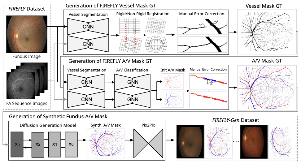 Overview of the FIREFLY (Fundus Images REgistered with FLuorescein angiographY) dataset construction process, used to train the deep learning based vessel segmentation and A/V classification modules.
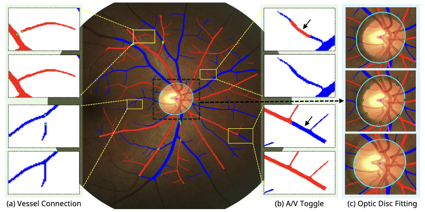 Examples of interactive semi-automatic editing tools for (a) vessel connection, (b) artery/vein label toggling of connected components, and (c) modification of the fitted optic disc ellipse.
 Overview of the vessel mask refinement and parameterization process. The vessel mask consists of two channels representing arteries and veins.
Why it matters
Published in Scientific Reports, this work offers a practical clinical tool that balances automation with expert oversight, directly improving the reliability of retinal vessel measurements used in ophthalmic diagnosis.