A novel vector field analysis for quantitative structure changes after macular epiretinal membrane surgery
Published in Scientific Reports, 2024
Authors: Seok Hyun Bae, Sojung Go, Jooyoung Kim, Kyu Hyung Park, Soochahn Lee, Sang Jun Park
TL;DR: A novel vector field analysis method to quantitatively measure structural changes in the retina before and after macular epiretinal membrane surgery.
What this paper is about
Epiretinal membrane (ERM) surgery aims to restore macular structure, but quantifying exactly how the retinal anatomy changes after surgery has been challenging. Traditional metrics like thickness maps miss the spatial deformation patterns that reveal how the retina recovers.
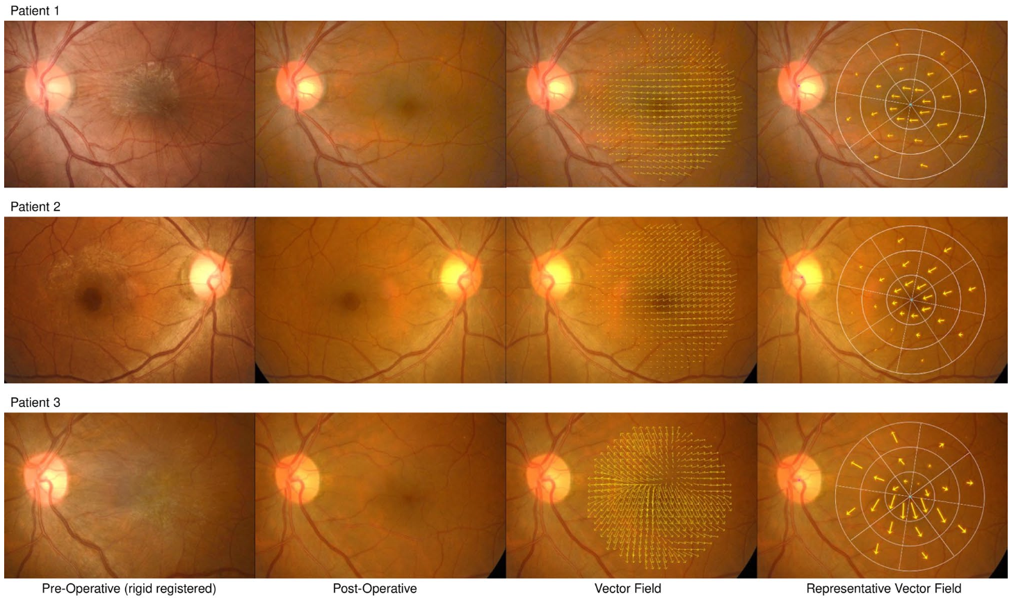 Representative retinal fundus images and visualized displacement vectors after rigid registration. Preoperative (first column) and postoperative 22 months (second column) retinal fundus images after rigid registration in reference to the postoperative images from three patients who underwent epiretinal membrane removal. In the third column, after calculation of pixel-wise retinal displacement of preoperative images in reference to postoperative image, the displacement vectors were visualized in every 25 pixels of presented postoperative retinal fundus images. The summed vectors of each 24 sectors are indicated by arrows in the fourth column. Note that a horizontally flipped image was used for the right eye of patient 2 (second row).
Key idea
The authors propose a vector field analysis framework that captures and quantifies the directional structural changes in the macula following ERM surgery. This provides a richer, spatially-aware representation of how retinal layers shift and recover compared to simple scalar measurements.
Why it matters
This gives ophthalmologists a more precise tool for evaluating surgical outcomes, potentially guiding post-operative care and helping predict visual recovery after ERM surgery.