Fully automatic 3-D segmentation of knee bone compartments by iterative local branch-and-mincut on MR images from osteoarthritis initiative (OAI).
Published in IEEE International Conference on Image Processing (ICIP), 2009
Authors: Sang Hyun Park, Soochahn Lee, Hackjoon Shim, Il Dong Yun, Sang Uk Lee, Kyoung Ho Lee, Heung Sik Kang, Joon Koo Han
TL;DR: A fully automatic method for 3D segmentation of knee bone compartments in MR images using an iterative local branch-and-mincut algorithm, requiring no manual initialization.
What this paper is about
Segmenting bone structures in knee MRI is important for orthopedic diagnosis and surgical planning, but most methods at the time required manual seed placement or other user interaction. Fully automatic approaches were needed to handle the large datasets coming from the Osteoarthritis Initiative.
Key idea
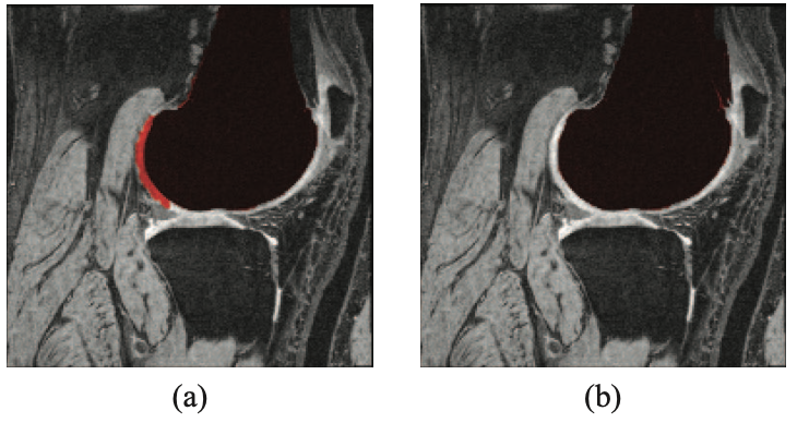 Comparison between results obtained by applying (a) shape priors only and (b) combined shape and intensity priors for an abnormal case (#1) where (a) shows better DSC perfor- mance. Despite the higher DSC, the result of (b) is preferable to (a), due to critical error, i.e., the misclassification of cartilage as bone.
The paper proposes an iterative local branch-and-mincut approach that segments knee bone compartments in 3D MR images without any manual intervention. By iteratively refining the segmentation through local graph-cut operations, the method achieves robust results even in areas with ambiguous boundaries.
Why it matters
Full automation removes the bottleneck of manual interaction, making it practical to process hundreds or thousands of knee MRI scans for large-scale clinical studies on osteoarthritis.