3-D segmentation of articular cartilages by graph cuts using knee MR images from osteoarthritis initiative.
Published in SPIE Medical Imaging, 2008
Authors: Hackjoon Shim, Soochahn Lee, Bohyeong Kim, Cheng Tao, Samuel Chang, Il Dong Yun, Sang Uk Lee, C. Kent Kwoh, Kyongtae Ty Bae
TL;DR: This paper applies graph cuts to automatically segment articular cartilage in 3D knee MR images from the Osteoarthritis Initiative, enabling quantitative cartilage analysis for osteoarthritis research.
What this paper is about
Osteoarthritis is one of the most common joint diseases, and tracking cartilage loss over time in MRI scans is critical for diagnosis and treatment monitoring. However, manually segmenting cartilage in 3D MR images is extremely time-consuming and prone to inter-observer variability.
Key idea
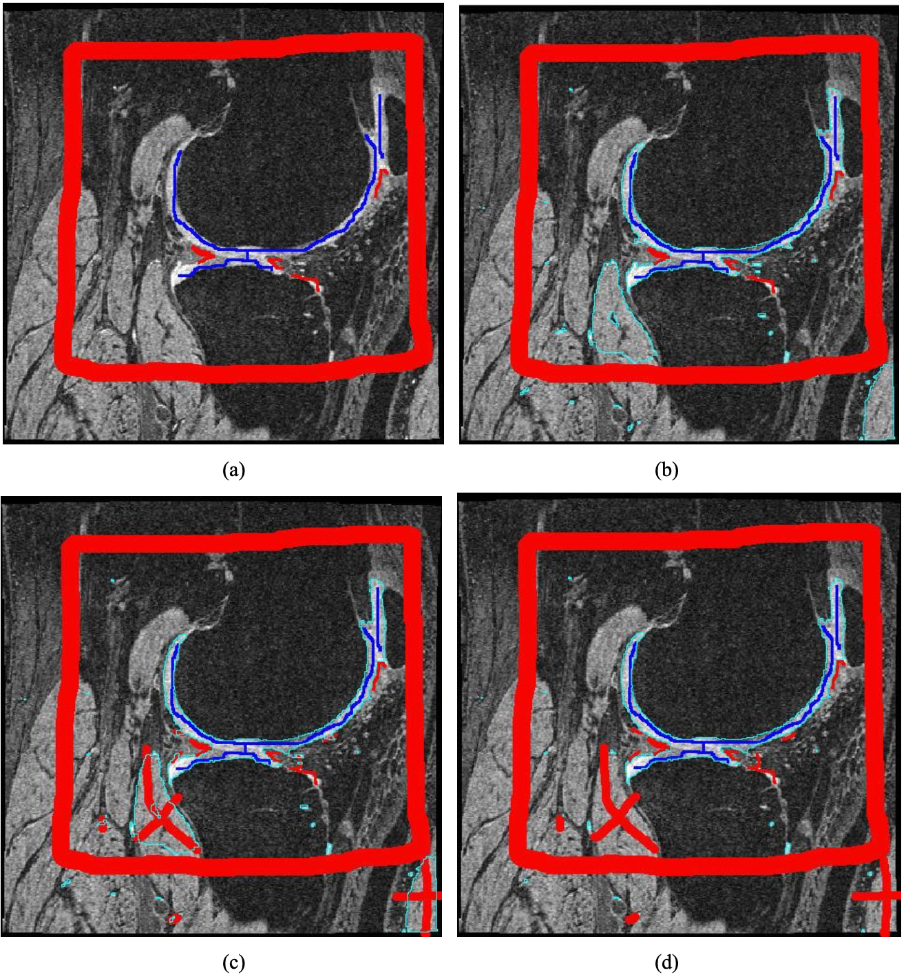 Cartilage segmentation on a slice using an s/t graph cut algorithm with hard constraints: (a) initial seeds, (b) segmentation using hard constraints of (a), (c) editing of the result (b), and (d) the final result.
The paper uses a graph-cut optimization framework to perform 3D segmentation of articular cartilages in knee MR images sourced from the Osteoarthritis Initiative (OAI). Graph cuts allow globally optimal segmentation by formulating the problem as an energy minimization over a voxel graph, balancing data fidelity with spatial smoothness.
Why it matters
By automating cartilage segmentation on OAI data, this work supports large-scale longitudinal studies of osteoarthritis progression, which would be infeasible with manual annotation alone.