A novel cascade classifier for automatic microcalcification detection.
Published in PLoS ONE, 2015
Authors: Seung Yeon Shin, Soochahn Lee, Il Dong Yun, Ho Yub Jung, Yong Seok Heo, Sun Mi Kim, Kyoung Mu Lee
TL;DR: A novel cascade classifier architecture for automatically detecting microcalcifications in mammograms, improving both detection sensitivity and computational efficiency.
What this paper is about
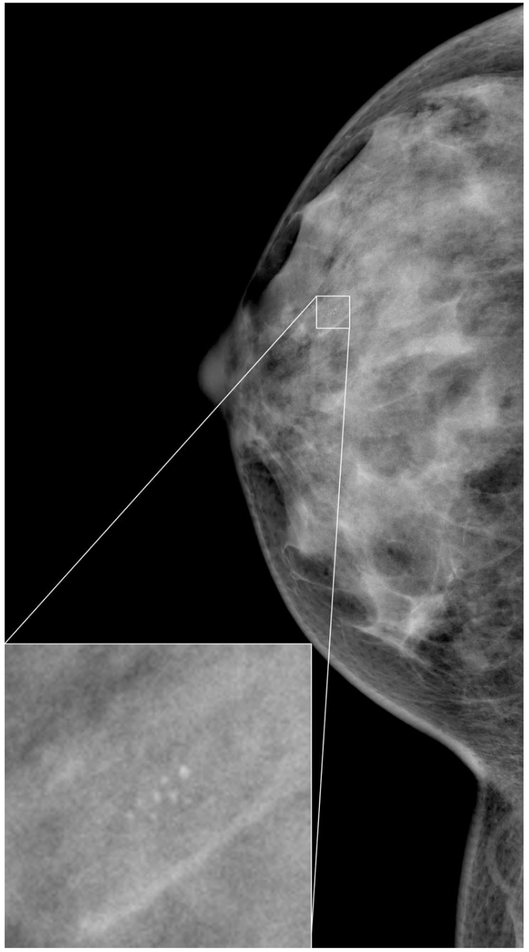 An example mammogram and the enlarged view of a contained microcalcification cluster.
Microcalcifications are tiny calcium deposits in breast tissue that can be early signs of cancer. Detecting them automatically in mammograms is hard because they are small, low-contrast, and easily confused with noise or other structures.
Key idea
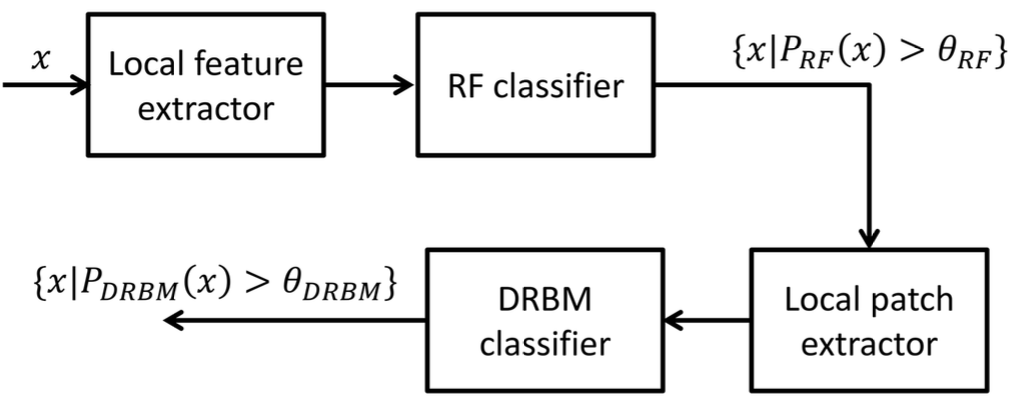 Schematic diagram of the proposed cascaded classification framework. Extracted local features from x, which are all pixels in a region of interest (ROI), are classified by the first RF classifier to produce μC probabilities $P_{RF}(x)$ and $\mu C$ candidates are detected by thresholding these probabilities with a certain value θRF. The $\mu C$ candidates are classified again by the second more sophisticated DRBM classifier. The input of the DRBM classifier is a local image patch and final detection results are again acquired by thresholding the probabilities $P_{DRBM}(x)$ with a certain value $\theta_{DRBM}$.
The paper designs a cascade classifier that progressively filters out non-calcification regions at each stage, allowing later stages to focus computational effort on the most promising candidates. This cascade structure balances high sensitivity (catching real calcifications) with low false positive rates by using increasingly sophisticated classifiers at each level.
Why it matters
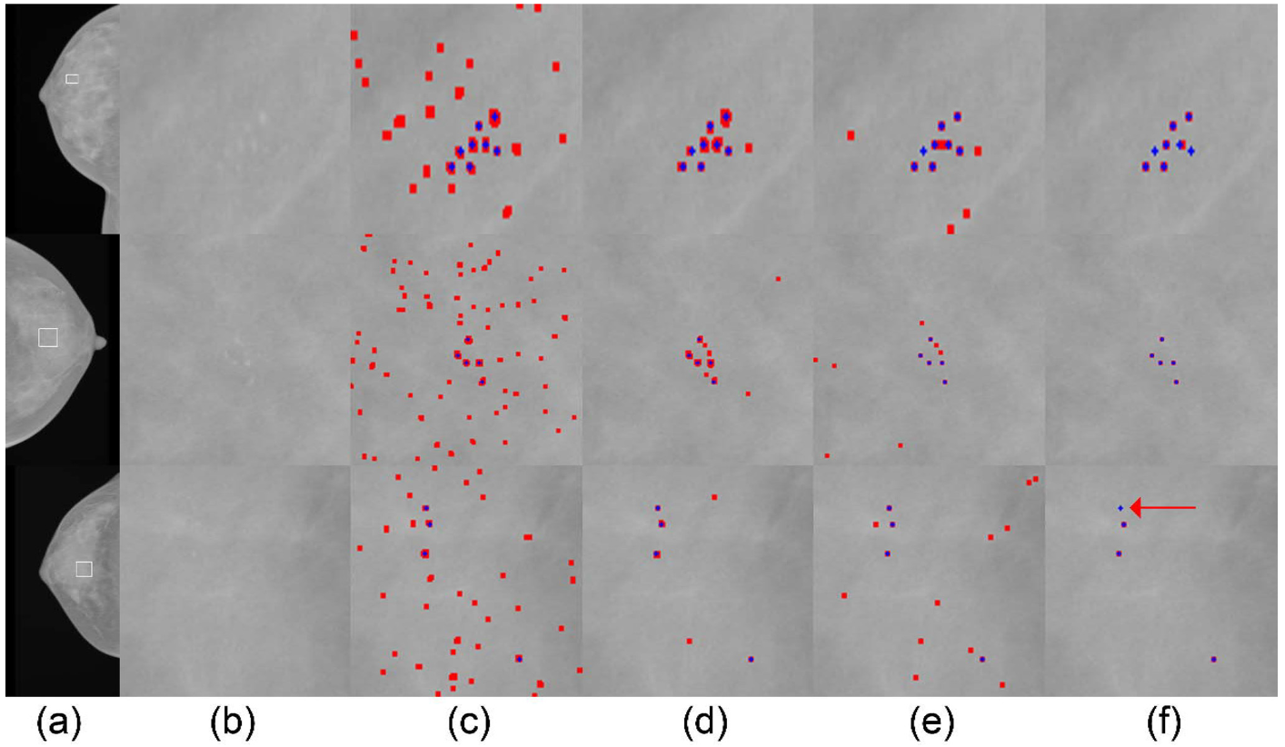 Qualitative results on the individual μC detection in the SNUBH-MDB-μCi. Each row represents a different case and each column shows (a) Test images, (b) Expanded views of ROIs (bounded by white boxes in (a)) containing annotated μCs and corresponding detection results by the methods of (c) Oliver et al. [13], (d) Wei et al. [15], (e) Ours (RF), (f) Ours (RF+DRBM). Blue crosses are annotated individual μCs and red pixels in the detection results are detected points by the methods.
The cascade design achieves strong detection performance while remaining computationally efficient, making it practical for integration into clinical computer-aided detection systems for breast cancer screening.