npj Digital Medicine · 2026

Interpretable Multimodal Retrieval Augmented Diagnosis for Breast Ultrasound with Multinational Clinical Validation and Reader Study

B-RAD reads like a radiologist: it retrieves BI-RADS-matched exemplars, localizes the lesion, and decides the category.

Han Jang1,2,† Subin An3,† Sunghyun Jung2 Hye Ji2 Miran Kim4 Soochahn Lee3,* Kyu Sung Choi2,5,6,* Su Min Ha2,5
1Interdisciplinary Program in Bioengineering, Seoul National University 2Department of Radiology, Seoul National University Hospital 3School of Electronic Engineering, Kookmin University 4Department of Radiology, Kyungpook National University Chilgok Hospital 5Department of Radiology, Seoul National University College of Medicine 6Healthcare AI Research Institute, Seoul National University Hospital
† Equal contribution  ·  * Corresponding authors
TL;DR — Paired ultrasound–report data are scarce, so B-RAD learns a BI-RADS-aware image–text space from unpaired data, uses retrieved exemplars to guide few-shot lesion localization, and steers the classifier toward the lesion with training-free foveal attention. Without fine-tuning, it reaches a biopsy-triage AUROC of 0.952 on an independent hospital cohort, and in a four-reader study it raises accuracy and inter-reader agreement.
Figure 1. What a reader sees with B-RAD. For an input ultrasound image, the system generates a pseudo-report, retrieves similar references with known diagnoses, and shows localizations with BI-RADS predictions and their evidence. The demo plays automatically; click the highlighted panel to step through it, or open it full screen.
8,311
images from 11 cohorts
in 7 countries
0.952
biopsy-triage AUROC on an independent institutional cohort (no fine-tuning)
0.53→0.68
inter-reader weighted κ
(moderate → substantial)
+9–25pp
reader accuracy gain with B-RAD across all four readers

Abstract

Breast ultrasound is central to breast cancer diagnosis but suffers from substantial inter-observer variability in Breast Imaging Reporting and Data System (BI-RADS) assessment, and existing deep learning models remain opaque. Multimodal large language models promise interpretable report-based reasoning, yet scarce paired image-report data yields weak visual-semantic alignment. We propose B-RAD, a BI-RADS-aware retrieval-augmented diagnosis framework that mirrors the radiologist workflow of retrieving exemplars, localizing lesions, and determining the BI-RADS category. Its retrieval module learns BI-RADS-aware alignment from unpaired data by jointly minimizing cross-modal mismatch and ordinal prediction error. Retrieved exemplars guide few-shot detection of the lesion together with its margin and posterior acoustic features, and the detected region prompts a segmentation model whose mask drives training-free foveal attention for classification. We validated B-RAD on eleven cohorts from seven countries totaling 8311 images, spanning internal validation and three external cohorts including an independent institutional cohort. The full pipeline reached a biopsy triage AUROC of 0.952 on that institutional cohort without fine-tuning, outperforming existing vision-language models. In a four-reader study, B-RAD assistance improved accuracy, raised inter-reader agreement from moderate to substantial, and reduced missed malignancies across all readers. These findings show that retrieval-augmented diagnosis can narrow the expertise gap and support accessible breast cancer screening in resource-limited settings.

Method

A cognition-aligned pipeline that follows the radiologist's reading order: retrieve, localize, then classify.
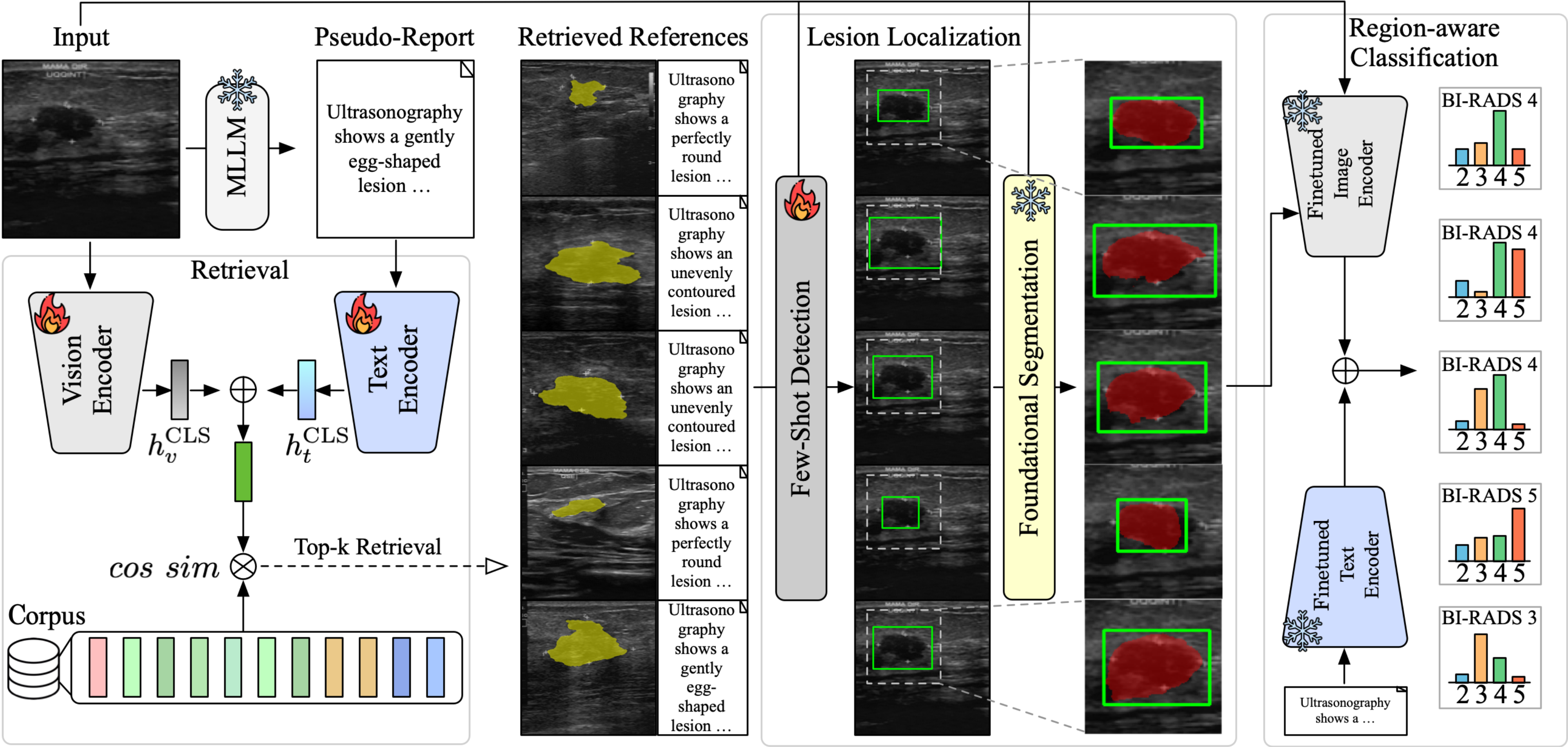
B-RAD pipeline: retrieval, few-shot detection and foundational segmentation, region-aware classification
Figure 2. Overview of B-RAD. An input image and an MLLM-generated pseudo-report query a BI-RADS-aware corpus. The retrieved exemplars guide few-shot lesion detection, the detected box prompts a segmentation foundation model, and the resulting ROI is injected into the vision encoder for region-aware BI-RADS classification.
1

BI-RADS-aware retrieval

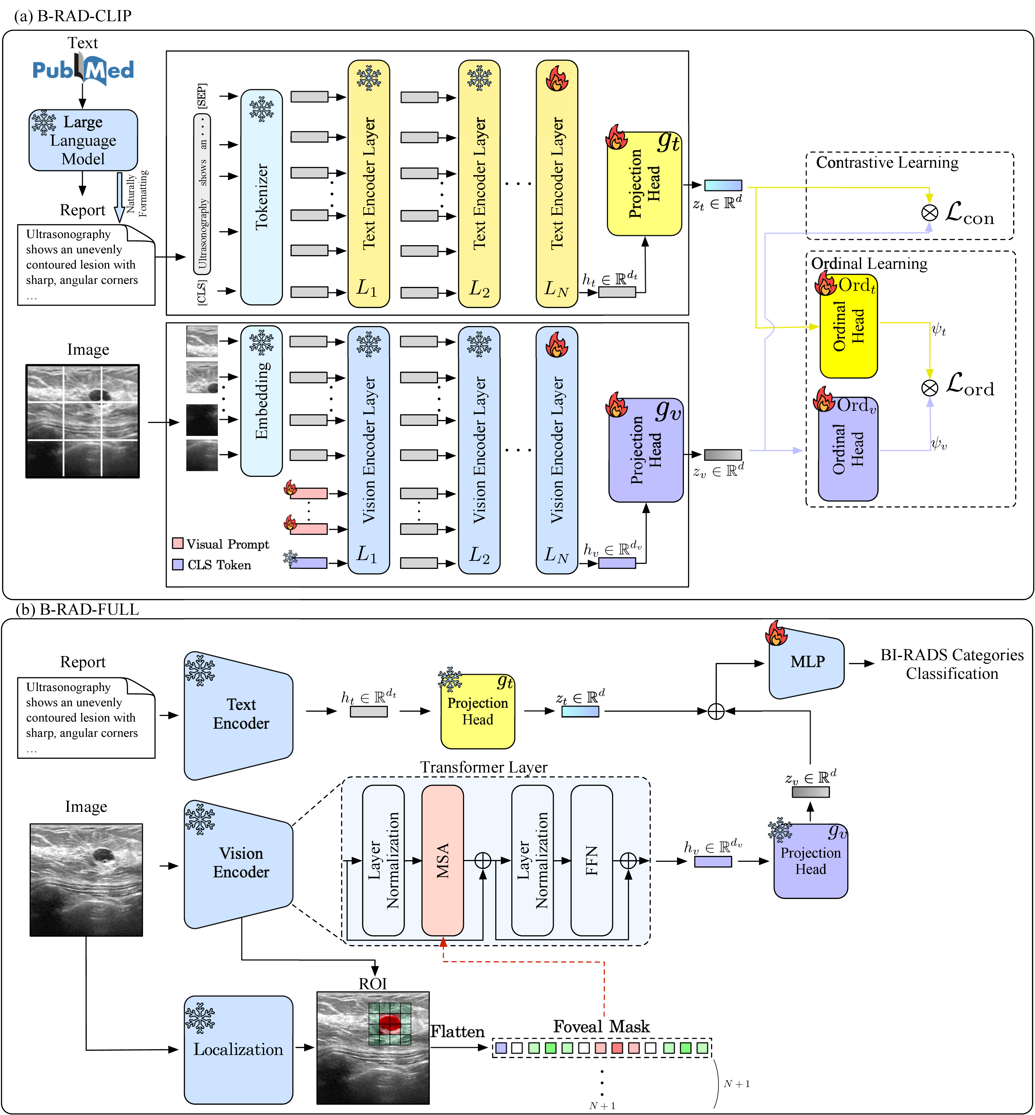
B-RAD-CLIP aligns images and structured lexicon text with a contrastive loss plus an ordinal loss over BI-RADS categories, so similar-looking cases are also similar in category. It is trained without paired image–report data.

B-RAD-CLIP
2

Exemplar-guided localization

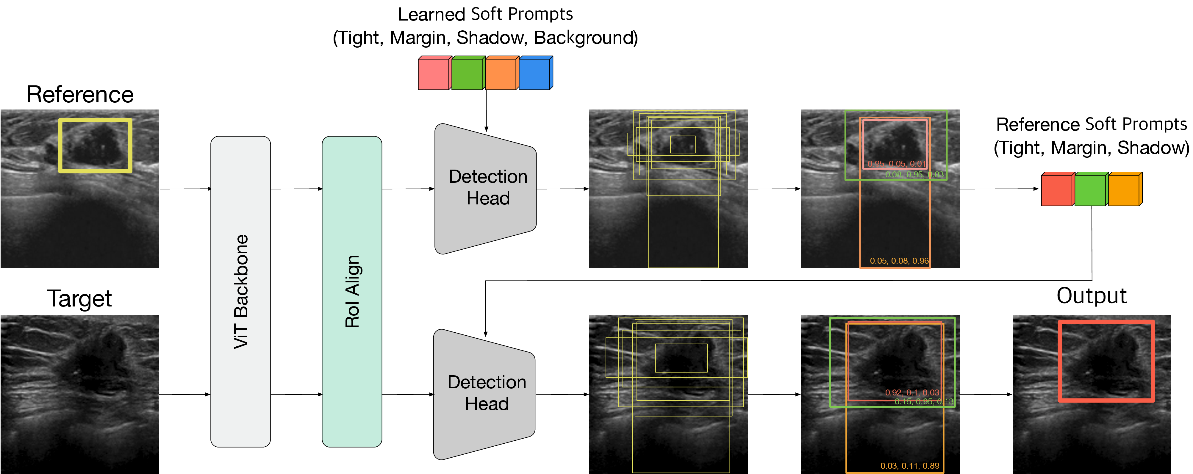
The top-k retrieved references serve as support examples for few-shot detection of the lesion along with its margin and posterior acoustic features. The predicted box then prompts a segmentation foundation model.

B-RAD-Det · B-RAD-Seg
3

ROI-guided classification

A training-free foveal attention mask built from the segmented ROI focuses the vision encoder on the lesion, and image, text, and ROI features are fused to predict BI-RADS 2–5.

B-RAD-FULL
Architecture details
Architecture of B-RAD-CLIP and B-RAD-FULL
B-RAD-CLIP and B-RAD-FULL. (a) Contrastive and ordinal learning with visual prompts. (b) ROI-derived foveal mask injected into multi-head self-attention, fused with text features for classification.
Retrieval-guided few-shot detection module
Retrieval-guided few-shot detection. Soft prompts learned from the reference (tight box, margin, shadow, background) condition the detection head on the target image.

Quantitative Results

B-RAD-FULL is the best model on every cohort, and the gap over off-the-shelf vision-language models widens on the independent hospital data.

ModelSettingAccF1AUROC [95% CI]
BI-RADS 5 vs othersBiopsy triage (2/3 vs 4/5)4-class weighted
OpenCLIPImg, Txt0.37 ± 0.030.41 ± 0.030.729 [0.667–0.787]0.799 [0.735–0.863]0.605 [0.568–0.644]
BiomedCLIPImg, Txt0.33 ± 0.030.36 ± 0.030.760 [0.701–0.812]0.890 [0.840–0.934]0.660 [0.620–0.699]
BMC-CLIPImg, Txt0.38 ± 0.030.30 ± 0.030.629 [0.566–0.694]0.671 [0.583–0.756]0.585 [0.528–0.638]
B-RAD-CLIPImg, Txt0.63 ± 0.030.63 ± 0.030.806 [0.750–0.855]0.951 [0.922–0.973]0.808 [0.772–0.843]
B-RAD-FULLImg, Txt, ROI0.63 ± 0.030.64 ± 0.030.807 [0.757–0.855]0.952 [0.925–0.973]0.809 [0.774–0.843]

n = 289, BI-RADS 2–5, collected from routine diagnostic breast ultrasound at a tertiary referral hospital (83.7% BI-RADS 4–5). All models were trained only on public ID cohorts and evaluated without fine-tuning.

Qualitative Results

Every prediction comes with retrieved evidence, a localized lesion, and attention that can be inspected.

BI-RADS-aware retrieval

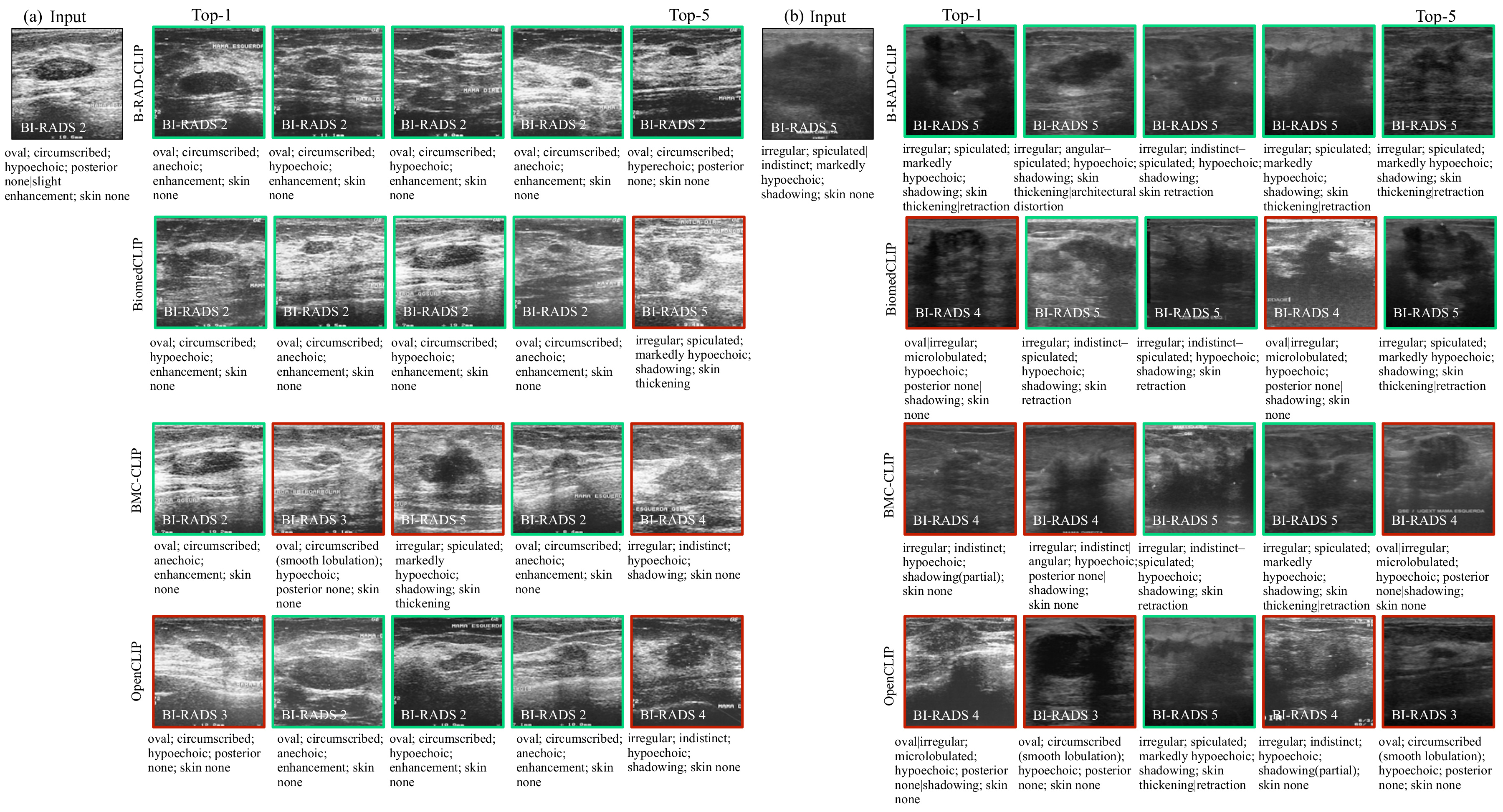
Top-5 retrieved exemplars for a BI-RADS 2 and a BI-RADS 5 query
Figure 4. Top-5 retrieved exemplars for (a) a benign BI-RADS 2 query and (b) a malignant BI-RADS 5 query. Green frames match the query category; red frames do not. Baselines often retrieve exemplars from the wrong category.

Lesion localization

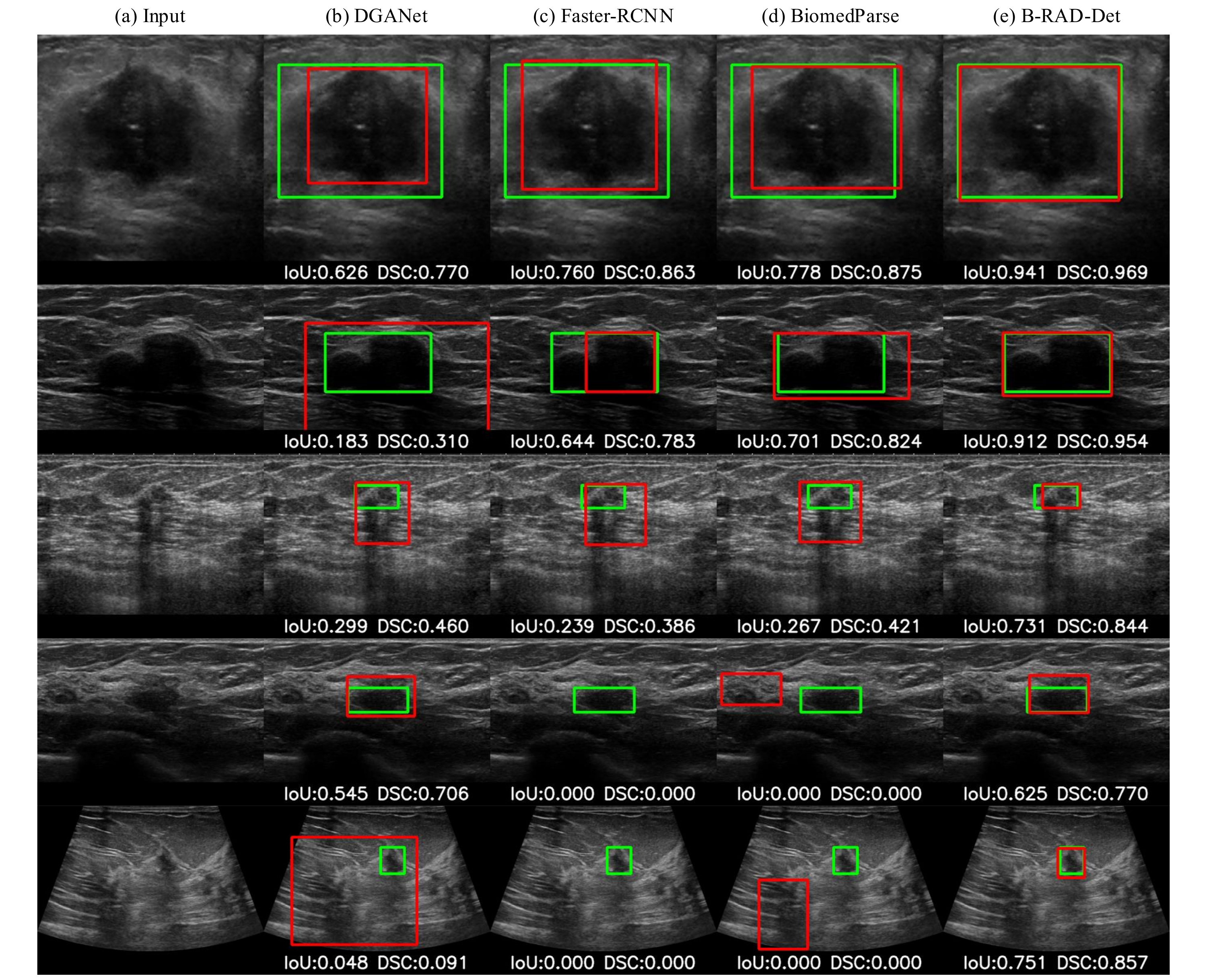
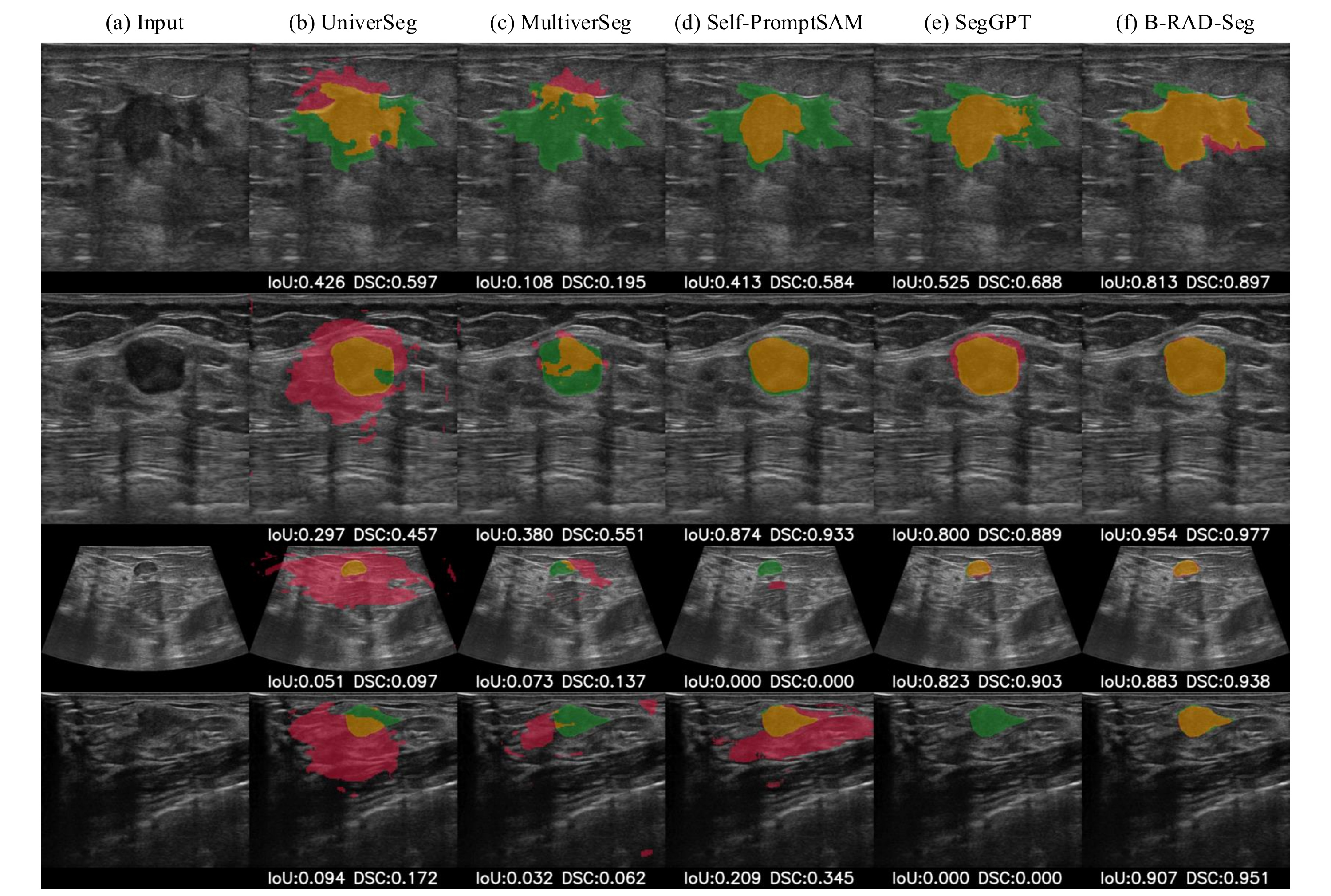
Few-shot lesion detection compared with baselines
Figure 6. Few-shot detection. Ground-truth boxes in green, predictions in red.
Few-shot segmentation masks compared with baselines
Figure 7. Segmentation masks from boxes predicted by B-RAD-Det, compared with in-context segmentation baselines.

Diagnostic evidence

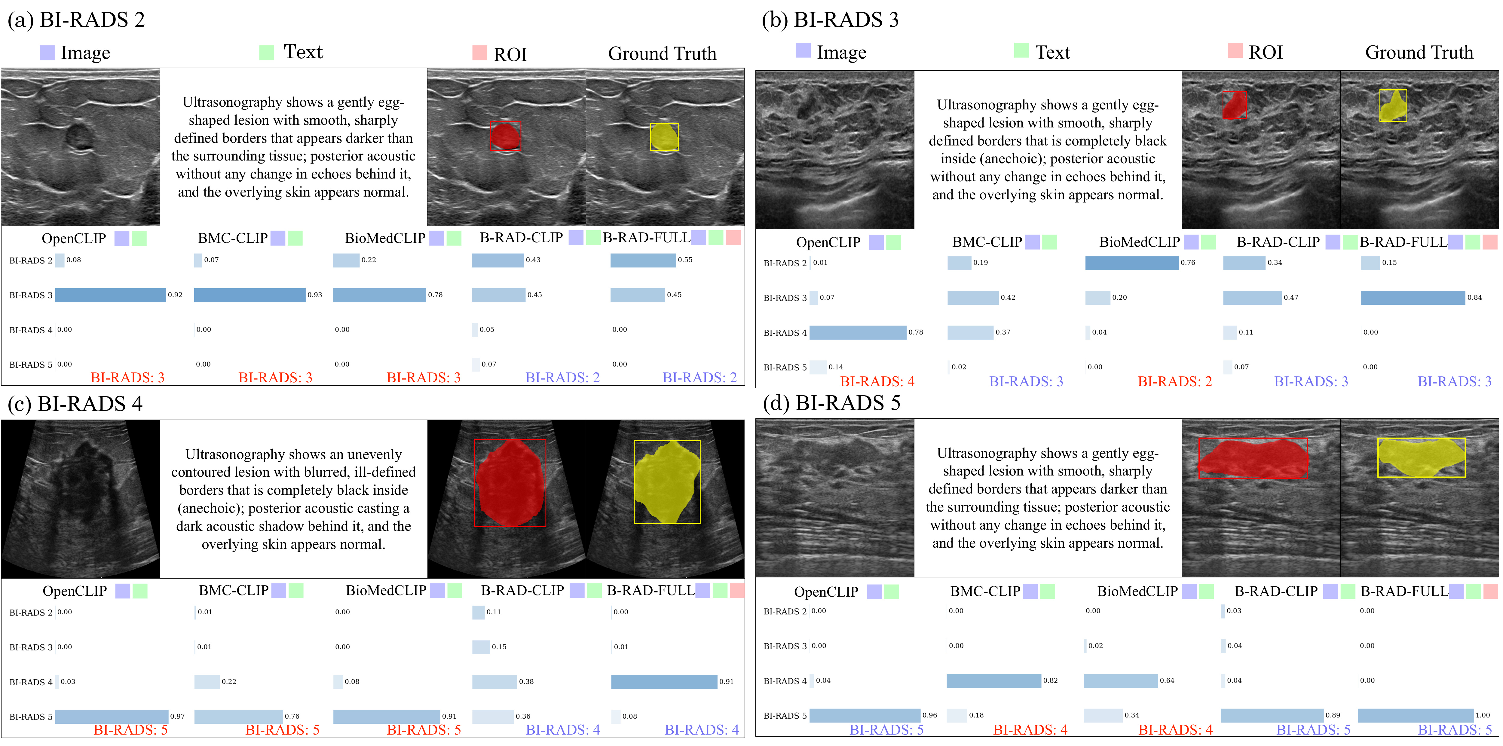
Representative cases for BI-RADS 2 to 5 with predicted probabilities by model
Figure 8. Representative test cases for BI-RADS 2–5 with the MLLM-generated description, predicted ROI (red), ground truth (yellow), and predicted class probabilities for each model.

Reader Study

Four radiologists with 2–5 years of experience each read 100 cases (65 benign, 35 malignant by pathology) unassisted, with an MLLM (MedGemma-4B), and with B-RAD.

Diagnostic accuracy (4-class BI-RADS)

Agreement with the expert BI-RADS assessment, %
Baseline+ MLLM+ B-RAD

McNemar (Bonferroni α = 0.017): Baseline vs B-RAD p < 0.001 for Senior, Junior and Board-certified readers; PGY-2 p = 0.136. MLLM assistance showed no significant improvement for any reader (all p > 0.4).

Inter-reader agreement

Quadratic-weighted κ across 4 readers
0.534
Baseline · moderate
→
0.682
+ B-RAD · substantial
Condition% AgreeBiopsy κFleiss' κ
Baseline50.30.3990.292
+ MLLM54.50.4900.306
+ B-RAD62.70.5760.463

Missed malignancies (biopsy decision)

% of malignant cases rated BI-RADS 2/3 (lower is better)
ReaderBaseline+ MLLM+ B-RAD
Senior breast (5 yr)14.317.111.4
Junior breast (4 yr)11.425.78.6
Board-certified (4 yr)37.128.617.1
PGY-2 resident (2 yr)25.737.111.4

Subjective evaluation (5-point Likert, mean of 4 readers)

Dimension+ MLLM+ B-RAD
Diagnostic confidence3.373.77
Interpretability2.994.00
Clinical utility2.913.74

All three readers with less experience exceeded the unassisted senior breast radiologist (54.0%) when assisted by B-RAD.

Appendix

Additional results from the paper. Figure numbers follow the article.

Multinational cohorts 11 datasets · 7 countries · 8,311 images

Six in-distribution datasets for training, four public external cohorts, and one independent Korean hospital cohort that was never used for training.

SetDatasetImagesCountryCenterAnnotationsUsageBI-RADS 2 / 3 / 4 / 5
In-distribution (ID)
IDHiSBreast287VietnamSingleBI-RADSCls30 / 77 / 69 / 111
IDOASBUD (RF)200PolandSingleBI-RADSCls0 / 62 / 88 / 50
IDBUS-BRA1,875BrazilSingleBI-RADS, MaskCls, Det/Seg562 / 463 / 693 / 157
IDBUSI-WHU927ChinaSingleMaskDet/Seg–
IDBUSI780EgyptSingleMaskDet/Seg–
IDBUS-UCLM264SpainSingleMaskDet/Seg–
Out-of-distribution (OOD)
OODBrEaST252PolandMultiBI-RADS, MaskCls, Det/Seg30 / 37 / 139 / 46
OODBUS-CoT3,232ChinaMultiBI-RADS, MaskCls, Det/Seg73 / 976 / 1942 / 241
OODBUS-STU42ChinaSingleMaskDet/Seg–
OODUDIAT163SpainSingleMaskDet/Seg–
Independent clinical validation
ClinicalInstitutional289South KoreaSingleBI-RADSCls19 / 28 / 127 / 115

Cls: classification, Det: detection, Seg: segmentation. Total 8,311 images (ID 4,333 · OOD 3,689 · Clinical 289).

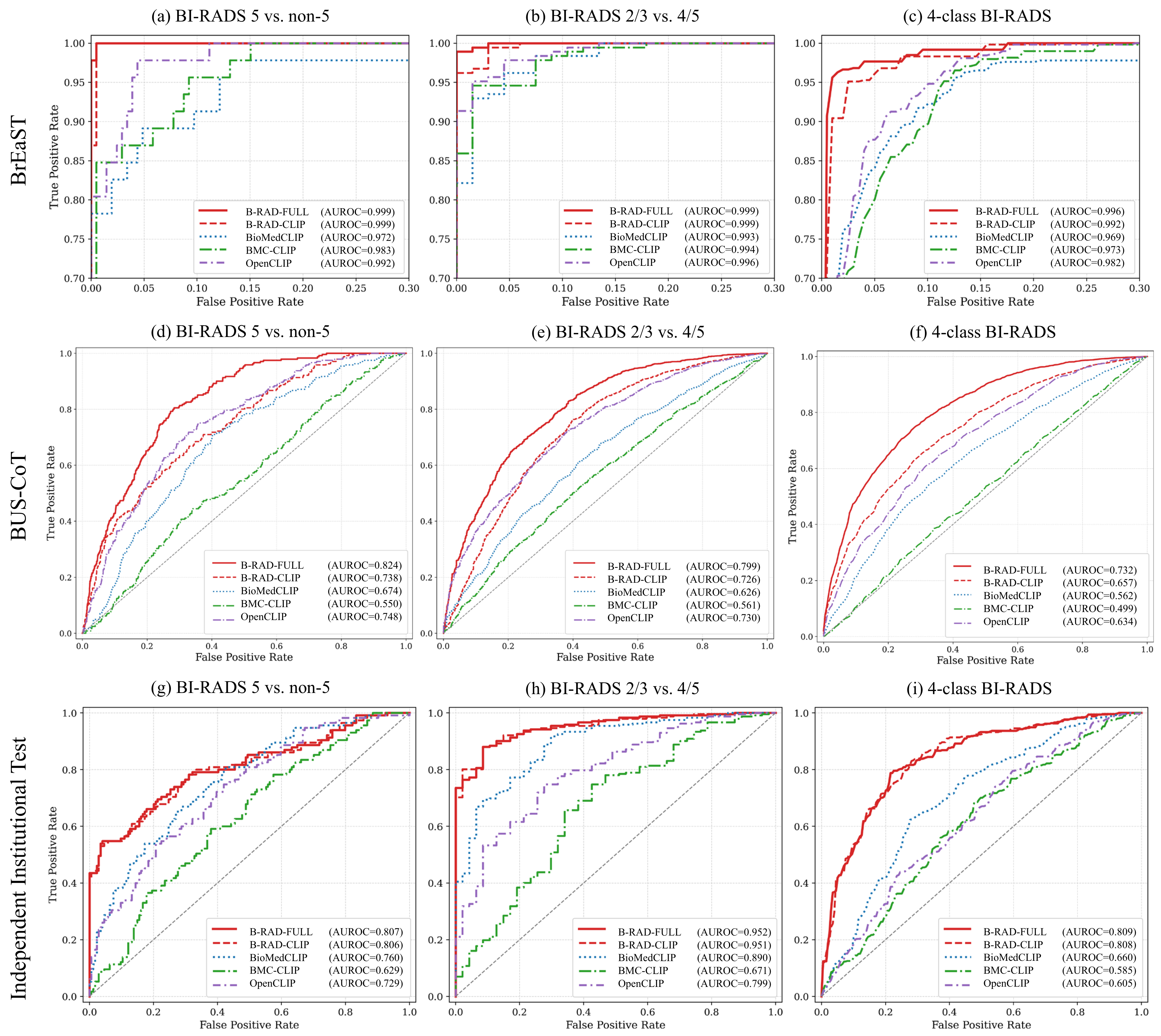
ROC curves Figure 3 · BrEaST, BUS-CoT and institutional cohorts
ROC curves on BrEaST, BUS-CoT and the institutional cohort
Figure 3. ROC curves on BrEaST (a–c), BUS-CoT (d–f), and the independent institutional cohort (g–i) at three clinical thresholds: BI-RADS 5 vs others, biopsy triage (BI-RADS 2/3 vs 4/5), and four-class one-vs-rest.
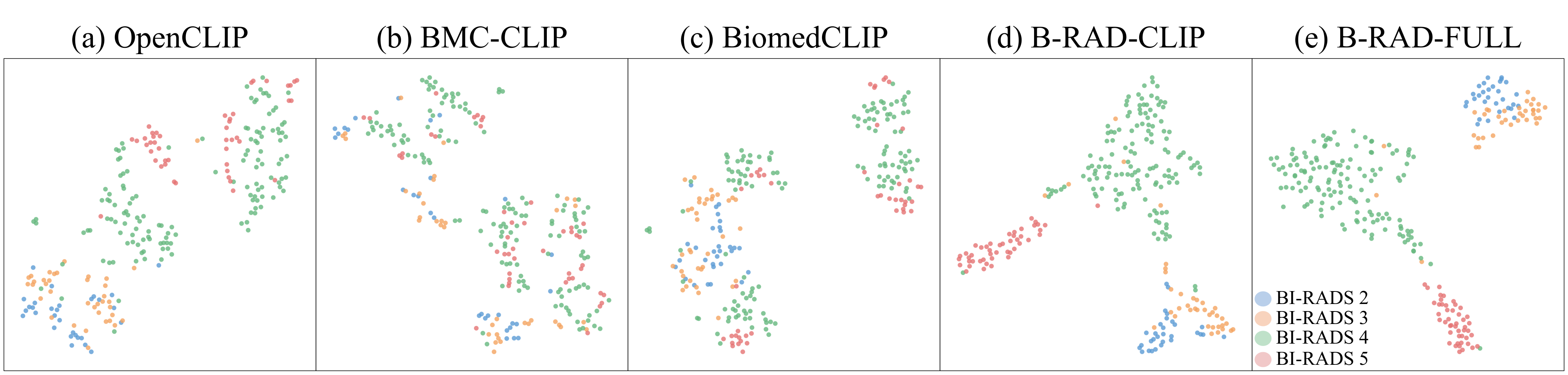
Embedding space Figure 5 · t-SNE of BI-RADS-aware embeddings
t-SNE of embedding spaces for baselines and B-RAD
Figure 5. t-SNE of the multimodal embedding space, colored by ground-truth BI-RADS. B-RAD-CLIP and B-RAD-FULL separate the categories, while baseline embeddings are entangled.
Attention analysis Figure 9 · how ROI injection shifts attention to the lesion
Attention maps before and after ROI injection, with attention ratio statistics
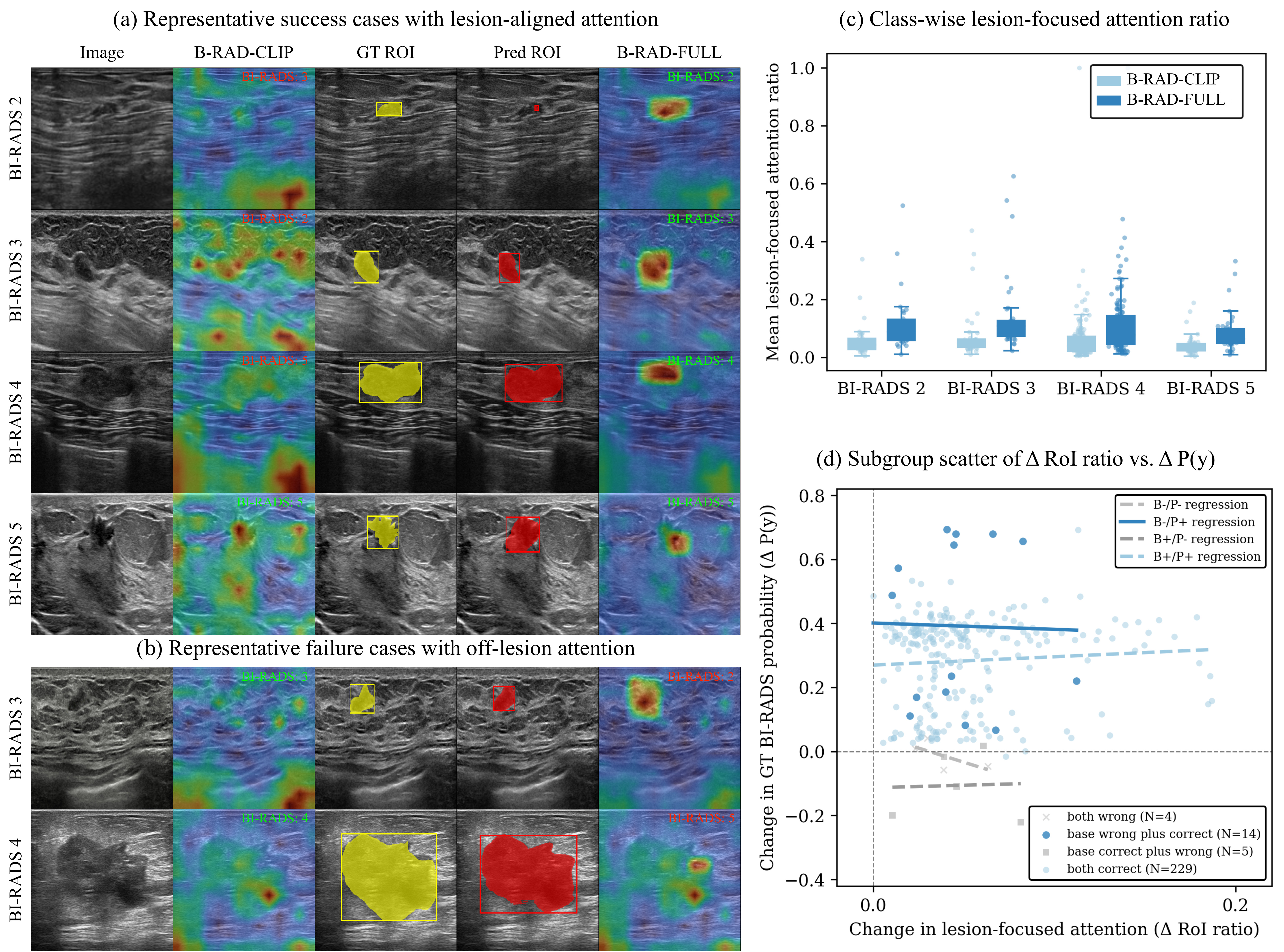
Figure 9. ROI injection shifts attention from diffuse patterns (B-RAD-CLIP) to the lesion (B-RAD-FULL), which corrects the BI-RADS prediction in success cases. Failure cases show attention that stays off target. The plots quantify the class-wise lesion-focused attention ratio and its relation to the change in ground-truth class probability.
Intended use. B-RAD is a research prototype for diagnostic decision support, applied after image acquisition as a second reader. It is not a medical device and is not intended for standalone clinical diagnosis.

BibTeX

If you find this work useful, please cite:

@article{jang2026brad,
  title   = {Interpretable multimodal retrieval augmented diagnosis for breast
             ultrasound with multinational clinical validation and reader study},
  author  = {Jang, Han and An, Subin and Jung, Sunghyun and Ji, Hye and
             Kim, Miran and Lee, Soochahn and Choi, Kyu Sung and Ha, Su Min},
  journal = {npj Digital Medicine},
  year    = {2026},
  doi     = {10.1038/s41746-026-03272-3}
}